⬆️
Multi-format upload
Upload standard images and whole-slide images including SVS, TIFF/TIF, PNG, JPG/JPEG, NDPI, SCN, MRXS, VMS/VMU and related formats.
Smart Pathology is an intelligent platform for pathology image upload, high-resolution viewing, AI-assisted analysis, structured reporting, and traceable analysis history — developed and implemented by Pooya Pardazan Asre Hooshmand.
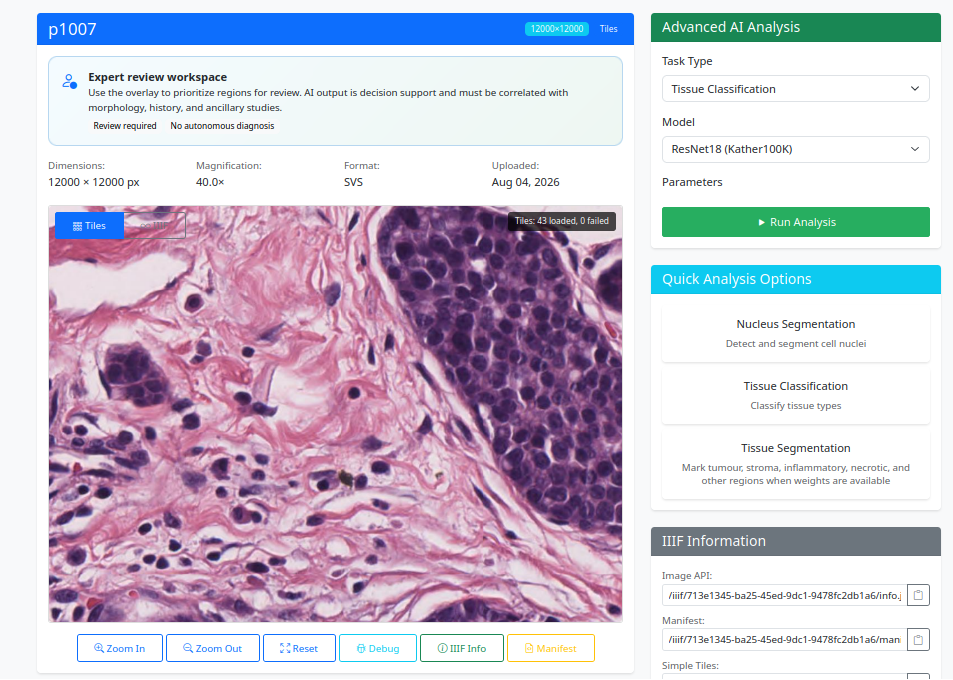 Real Smart Pathology interface
The workflow follows the real sequence of digital pathology work: upload the case, inspect the slide, select the analysis, run the model, review the overlays, generate the report, and revisit the analysis later.
Upload standard images and whole-slide images including SVS, TIFF/TIF, PNG, JPG/JPEG, NDPI, SCN, MRXS, VMS/VMU and related formats.
Navigate large images, inspect regions of interest, work with tiles, and review the slide at up to 40× magnification.
Choose tissue classification, tissue segmentation, or nucleus analysis, then select the appropriate model for the task.
Review visual overlays, quantitative findings, structured reports, and a persistent record of completed analyses.
These are real screenshots from the working Smart Pathology platform. The slideshow follows a case from the dashboard and upload page to model selection, AI execution, and final reports.
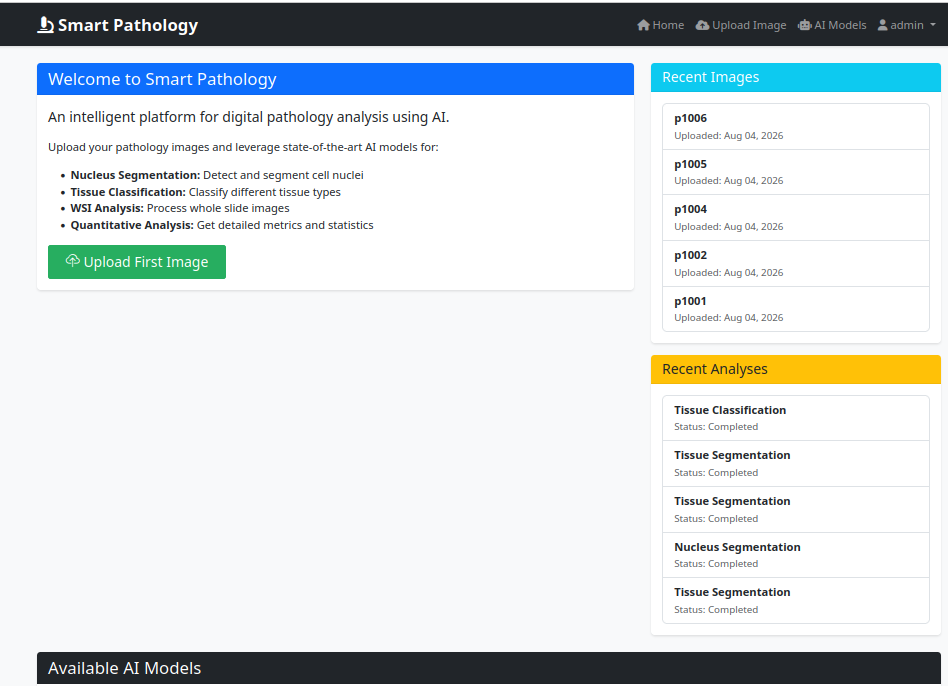
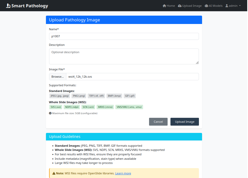
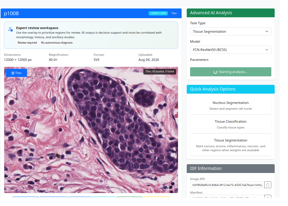
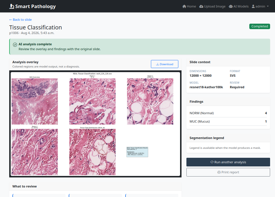
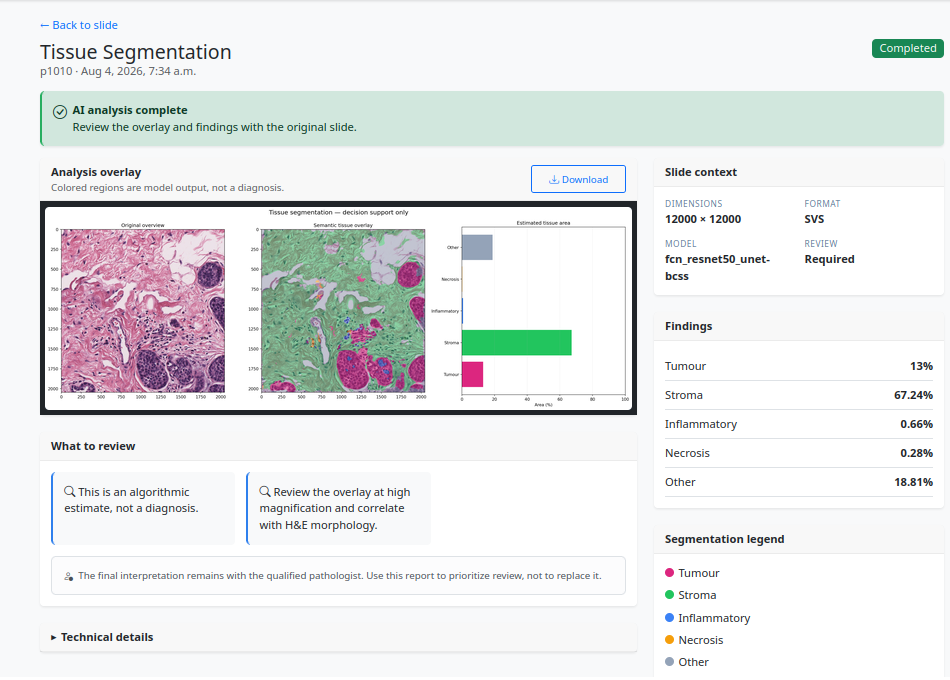
Image management, high-resolution viewing, analysis orchestration, AI execution, result composition, reporting, and history storage are separated into clear functional stages.
Attach the pathology image to a named case.
Check format, metadata, dimensions, and WSI compatibility.
Render the image or slide using a tiled multi-resolution viewer.
Select classification, segmentation, or nucleus analysis and the related model.
Execute inference and model-specific post-processing.
Inspect overlays, tissue percentages, classes, cell information, and findings.
Store the report, outputs, model information, and complete analysis history.
Each model is presented with a dedicated pathology visual, the target output classes, and the relevant training-data context — similar to the model slides used in the presentation.
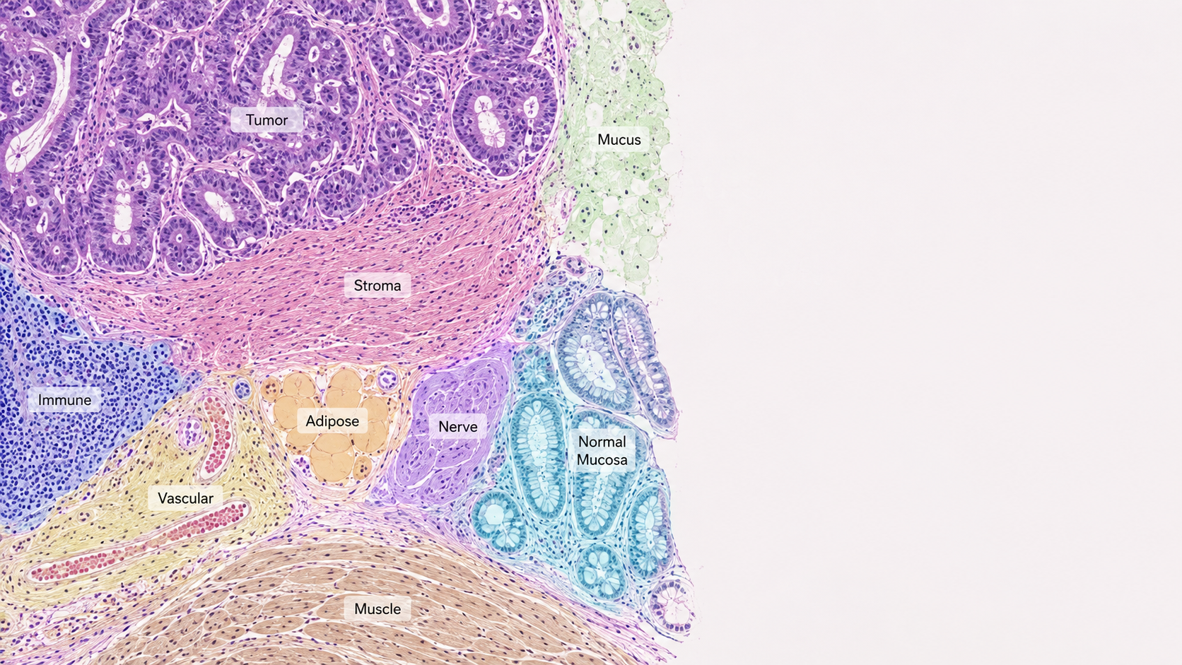
The classifier works on tissue patches and identifies the dominant colorectal tissue pattern. In the platform, these predictions can be used to build a regional tissue map and summarize the tissue composition of the selected image area.
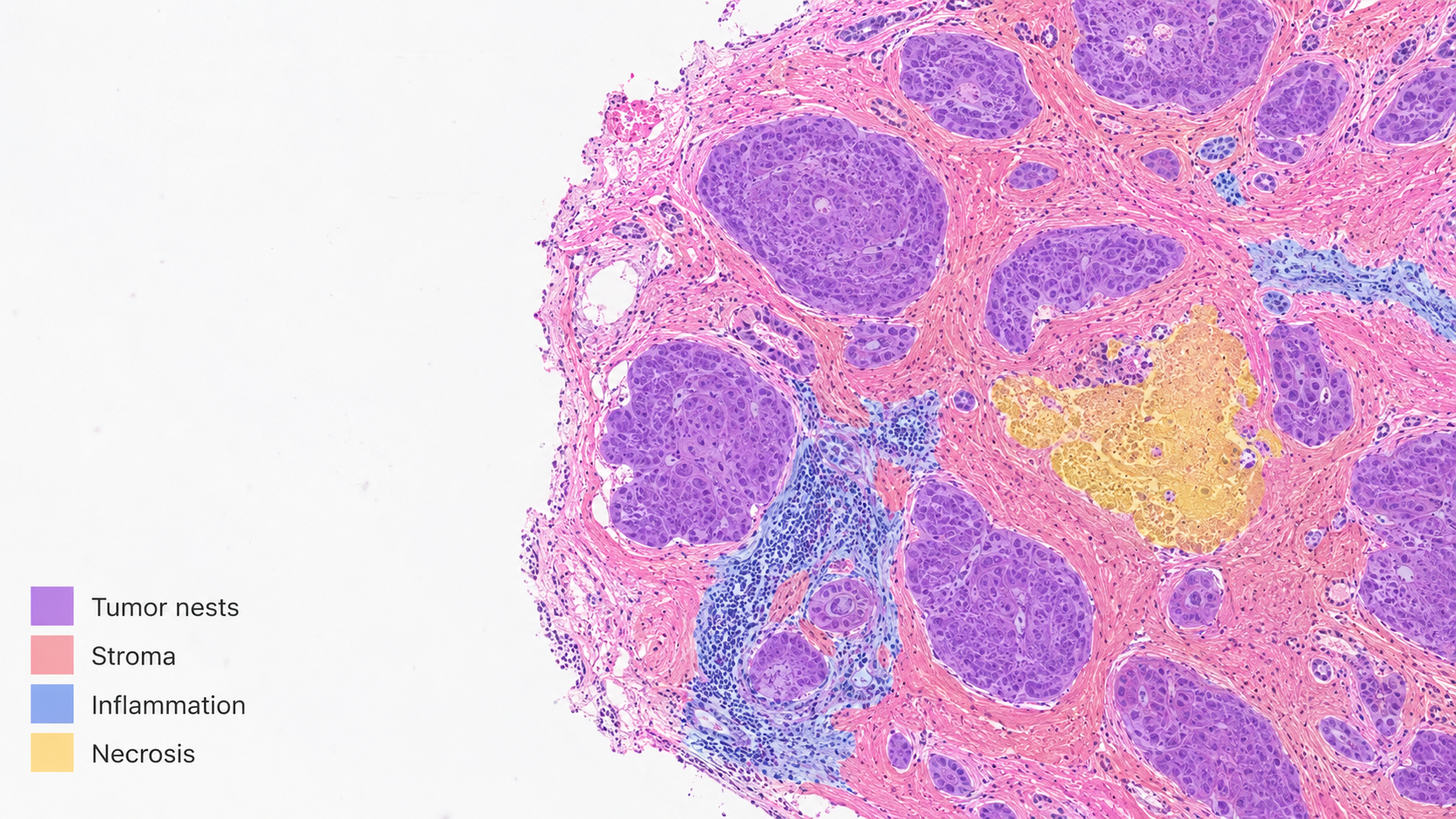
This model produces pixel-level tissue masks rather than a single label. The platform can render those masks directly over the original slide and calculate the approximate area and proportion of each tissue compartment.
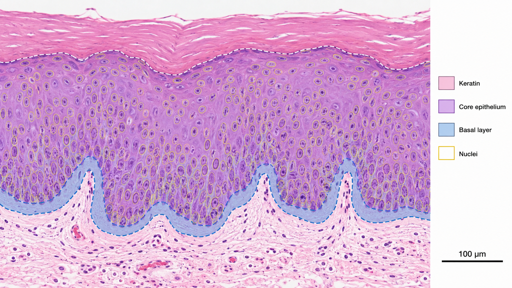
This model combines two levels of analysis: semantic segmentation of the oral epithelium and instance-level analysis of nuclei. This makes the output closer to pathology review, where both tissue architecture and cellular morphology are considered together.
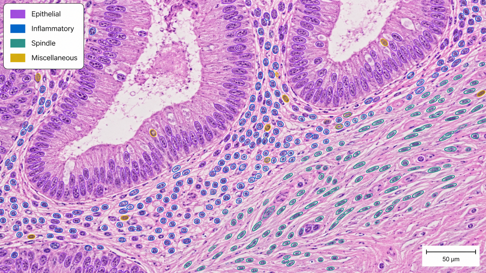
Instead of describing the tissue region, this model identifies individual nuclei, separates touching nuclei, and assigns them to clinically meaningful nucleus categories. The result can support cell counting, density analysis, and spatial characterization of the microenvironment.
The platform keeps the pathologist in control while making the AI result transparent, reviewable, quantitative, and easy to revisit.
Masks, nucleus contours, tissue labels, and color-coded regions are placed directly over the pathology image.
Tissue percentages, cell counts, class distributions, and other task-specific measurements are summarized for review.
The report combines slide metadata, selected model, analysis findings, technical context, and review guidance.
Completed analyses are retained so users can reopen the case, compare outputs, or generate the report again.
Smart Pathology is designed to support pathology specialists with AI-assisted visual evidence, quantitative information, and a reproducible digital workflow — while keeping expert interpretation at the center of the process.
See the Workflow